
Nvidia · Filed Jun 25, 2026 · Published Oct 29, 2026

Nvidia Files Patent for AI That Spots and Describes Disease in Medical Scans

Doctors' notes on X-rays, like 'diffused opacity in left-lung,' turn out to be free training material. Nvidia's patent filing uses them to teach software to describe what it sees.
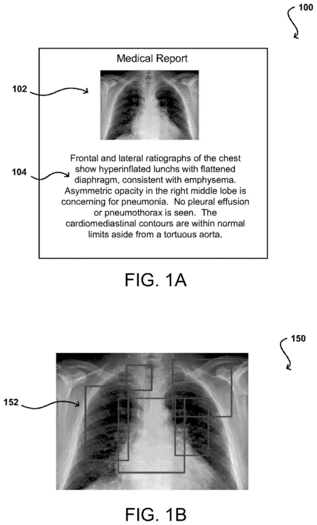
A chest X-ray report paired with highlighted regions that help an AI pinpoint signs of lung disease. Drawing from patent filing US 2026/0341060 A1.
A chest X-ray report paired with highlighted regions that help an AI pinpoint signs of lung disease.
See all 50 drawings from this filing ↓
Publication number US 2026/0341060 A1 US20260341060A1
Applicant NVIDIA Corporation
Filing date Jun 25, 2026
Publication date Oct 29, 2026
Inventors Daguang Xu, Xiaosong Wang, Lickkong Tam, Riddhish Bhalodia, Kevin Lu, Yuhong Wen, Ali Hatmizadeh
US classification 382/128
Status when we published Waiting for an examiner (Jul 15, 2026)
Parent application is a Continuation of 17410471 (filed 2021-08-24)
Document 20 claims

What Nvidia's X-ray reading AI actually does

Every time someone gets a chest X-ray, a doctor studies the image and types a short note, something like diffused opacity in left-lung. Those notes pile up by the thousands, sitting right next to the images they describe. Nvidia's patent filing treats that pile as a free teacher.

The software learns by pairing the pictures with key words from the notes, such as left, small, diffuse or severe. Once it is trained, it no longer needs a note at all. Hand it a new scan and it marks the suspicious areas and gives a probability for each descriptive word, a bit like a first-pass read of where the trouble is and how bad it looks.

Why bother? Hand-labeling medical images is slow and expensive, and the filing says that often means smaller training sets and less accurate software. Its main example is pneumonia, including cases tied to COVID-19.

From the filing · CLAIM 1
… transform the feature vector for the at least one candidate region of interest into at least one modified region feature vector based, at least in part, on a normalized representation of geometric information for a corresponding candidate region of interest; …

Translation: The system adjusts data about a specific area of the scan by factoring in its actual size and shape.

How the AI weighs and pools suspicious regions

Claim 1 describes a processor that takes one medical image and first finds candidate regions of interest, meaning boxes around spots that might matter. Each box gets a feature vector, which is a list of numbers summarizing what that patch of the image looks like.

Next comes the distinctive step. The feature vector is transformed using a normalized representation of the box's geometry, meaning the box's coordinates scaled to a standard range so its position and size are folded into the numbers. A dependent claim adds that the box's classification score is joined on too.

The regions then get weights (how much each one should count), and the weighted vectors are pooled into a single aggregate image representation. From that summary, the system outputs an attribute probability vector: a list of probabilities, one per descriptive term, that characterizes the object it found.

The description explains how the network learns. Training pairs chest X-rays with the text of radiology reports. A language tool pulls out a fixed set of attribute words (22 in one example, including left, bilateral, diffuse, moderate and severe), and a matching step teaches the network which regions fit which words. The example detector is a standard object-detection network. After training, only the image is needed.

From the filing · THE ABSTRACT
Apparatuses, systems, and techniques are presented to detect one or more objects in one or more images. In at least one embodiment, one or more neural networks can be used to detect one or more objects in one or more images based, at least in part, on textual descriptions of the one or more objects.

Translation: AI tools scan medical images to spot specific anomalies using text descriptions as a guide.

Why descriptive scores could matter in radiology

The practical payoff is richer output. Instead of one score saying pneumonia is probably present, a system built this way could mark where it sees trouble and attach probabilities for words such as small, bilateral or severe. The description calls COVID-19 an important use case and says the approach could extend to other diseases, and to organs, bones or tumors in CT, MRI and ultrasound images.

The training angle matters too. The filing says hand-labeling images takes experts a lot of time and money, which often shrinks training sets and hurts accuracy. Reusing reports that doctors already wrote sidesteps part of that cost. Keep in mind this is a patent application describing a research-style method; nothing in it says the technique is in a clinic or a product.

That makes this Nvidia's 53rd filing in AI training and infrastructure we've tracked since May, alongside one on storing video inside a model and one on hidden scene completion.

Editorial take

Claim 1 covers a processor that takes a medical image, finds candidate regions within it, adjusts each region's data based on its position, combines everything into a single summary, and outputs probabilities for descriptive labels. Nothing in that claim limits it to a specific disease, body part, or scanning technology, so the protected boundary stretches across virtually any medical imaging context.

That breadth has real consequences. Any tool following those same steps, even with different training data or different medical applications, would fall inside the claim.

The abstract's headline feature, training the system on written clinical reports rather than hand-labeled images, does not appear in claim 1 at all. The patent's practical reach is therefore narrower than its most compelling capability suggests, and that gap matters before drawing conclusions about what this filing actually controls.

Get our take in your Top Stories

Liked this breakdown? Add Patentlyze as a preferred source on Google, and our plain-English take shows up more often in your Top Stories the next time Nvidia patent news breaks.

There are more where this came from

We read every patent application Big Tech publishes and send you the ones worth knowing. Plain English, free, every week.

The drawings

50 drawing sheets from US 2026/0341060 A1 · click any drawing to enlarge

Matching lung X-ray regions to a doctor's diagnosis notes, drawing sheet 1 from US 2026/0341060 A1
Sheet 1 Matching lung X-ray regions to a doctor's diagnosis notes
How the system compares scan areas to written doctor descriptions, drawing sheet 2 from US 2026/0341060 A1
Sheet 2 How the system compares scan areas to written doctor descriptions
Comparing AI disease predictions with doctor labels on chest scans, drawing sheet 3 from US 2026/0341060 A1
Sheet 3 Comparing AI disease predictions with doctor labels on chest scans
Training the AI by checking scans against real diagnoses, drawing sheet 4 from US 2026/0341060 A1
Sheet 4 Training the AI by checking scans against real diagnoses
Finding and describing diseases using scans and doctor notes, drawing sheet 5 from US 2026/0341060 A1
Sheet 5 Finding and describing diseases using scans and doctor notes
Detecting objects in medical images using accompanying text, drawing sheet 6 from US 2026/0341060 A1
Sheet 6 Detecting objects in medical images using accompanying text
Processor parts that handle calculations for the AI model, drawing sheet 7 from US 2026/0341060 A1
Sheet 7 Processor parts that handle calculations for the AI model
Dedicated chip parts paired with memory to run AI faster, drawing sheet 8 from US 2026/0341060 A1
Sheet 8 Dedicated chip parts paired with memory to run AI faster
Show the other 42 sheets
How cloud data centers manage hardware to run medical AI, drawing sheet 9 from US 2026/0341060 A1
Sheet 9 How cloud data centers manage hardware to run medical AI
Internal parts of a computer that analyzes medical scans, drawing sheet 10 from US 2026/0341060 A1
Sheet 10 Internal parts of a computer that analyzes medical scans
Components inside a portable device that runs diagnostic software, drawing sheet 11 from US 2026/0341060 A1
Sheet 11 Components inside a portable device that runs diagnostic software
Multiple graphics chips linked together to process medical images, drawing sheet 12 from US 2026/0341060 A1
Sheet 12 Multiple graphics chips linked together to process medical images
A USB drive that runs AI tasks, drawing sheet 13 from US 2026/0341060 A1
Sheet 13 A USB drive that runs AI tasks
High-speed connections linking multiple main processors and graphics chips, drawing sheet 14 from US 2026/0341060 A1
Sheet 14 High-speed connections linking multiple main processors and graphics chips
Shared memory layout between a main processor and graphics accelerators, drawing sheet 15 from US 2026/0341060 A1
Sheet 15 Shared memory layout between a main processor and graphics accelerators
Alternate direct connections between main processor cores and accelerators, drawing sheet 16 from US 2026/0341060 A1
Sheet 16 Alternate direct connections between main processor cores and accelerators
Connecting graphics hardware directly to an app's memory space, drawing sheet 17 from US 2026/0341060 A1
Sheet 17 Connecting graphics hardware directly to an app's memory space
Virtual memory sharing managed under a system supervisor, drawing sheet 18 from US 2026/0341060 A1
Sheet 18 Virtual memory sharing managed under a system supervisor
Pooling system memory across multiple graphics chips, drawing sheet 19 from US 2026/0341060 A1
Sheet 19 Pooling system memory across multiple graphics chips
Combining graphics, video, and AI processors onto a single chip, drawing sheet 20 from US 2026/0341060 A1
Sheet 20 Combining graphics, video, and AI processors onto a single chip
Dividing graphics tasks among dedicated visual processing units, drawing sheet 21 from US 2026/0341060 A1
Sheet 21 Dividing graphics tasks among dedicated visual processing units
Coordinating multiple programmable shader cores on one chip, drawing sheet 22 from US 2026/0341060 A1
Sheet 22 Coordinating multiple programmable shader cores on one chip
Inside layout of a processor core running fast math tasks, drawing sheet 23 from US 2026/0341060 A1
Sheet 23 Inside layout of a processor core running fast math tasks
Grouping multiple computing clusters to speed up heavy AI workloads, drawing sheet 24 from US 2026/0341060 A1
Sheet 24 Grouping multiple computing clusters to speed up heavy AI workloads
Computer hardware linking graphics chips and memory to train AI, drawing sheet 25 from US 2026/0341060 A1
Sheet 25 Computer hardware linking graphics chips and memory to train AI
A chip built to run multiple calculations at once, drawing sheet 26 from US 2026/0341060 A1
Sheet 26 A chip built to run multiple calculations at once
Memory parts that temporarily hold image data inside the chip, drawing sheet 27 from US 2026/0341060 A1
Sheet 27 Memory parts that temporarily hold image data inside the chip
One processing block that handles textures and image math, drawing sheet 28 from US 2026/0341060 A1
Sheet 28 One processing block that handles textures and image math
Core calculation engines and fast memory within a graphics chip, drawing sheet 29 from US 2026/0341060 A1
Sheet 29 Core calculation engines and fast memory within a graphics chip
Linking four chips together to speed up scan analysis, drawing sheet 30 from US 2026/0341060 A1
Sheet 30 Linking four chips together to speed up scan analysis
Video parts that process incoming visuals for the screen, drawing sheet 31 from US 2026/0341060 A1
Sheet 31 Video parts that process incoming visuals for the screen
Microscopic layout of a main processor that organizes computing steps, drawing sheet 32 from US 2026/0341060 A1
Sheet 32 Microscopic layout of a main processor that organizes computing steps
A specialized AI processor built to train medical models, drawing sheet 33 from US 2026/0341060 A1
Sheet 33 A specialized AI processor built to train medical models
Brain-inspired circuits that send signals like human neurons, drawing sheet 34 from US 2026/0341060 A1
Sheet 34 Brain-inspired circuits that send signals like human neurons
Main computer parts that handle medical sensors and graphics, drawing sheet 35 from US 2026/0341060 A1
Sheet 35 Main computer parts that handle medical sensors and graphics
Multi-core processor layout with shared memory rings, drawing sheet 36 from US 2026/0341060 A1
Sheet 36 Multi-core processor layout with shared memory rings
Internal layout of a graphics processor core, drawing sheet 37 from US 2026/0341060 A1
Sheet 37 Internal layout of a graphics processor core
Execution blocks inside a graphics processor core, drawing sheet 38 from US 2026/0341060 A1
Sheet 38 Execution blocks inside a graphics processor core
Hardware units that carry out math and logic instructions, drawing sheet 39 from US 2026/0341060 A1
Sheet 39 Hardware units that carry out math and logic instructions
A computer chip built to run healthcare AI, drawing sheet 40 from US 2026/0341060 A1
Sheet 40 A computer chip built to run healthcare AI
Internal chip cluster that schedules tasks and runs graphics logic, drawing sheet 41 from US 2026/0341060 A1
Sheet 41 Internal chip cluster that schedules tasks and runs graphics logic
How the chip loads images into local memory, drawing sheet 42 from US 2026/0341060 A1
Sheet 42 How the chip loads images into local memory
Multiprocessor core that executes AI instructions across parallel units, drawing sheet 43 from US 2026/0341060 A1
Sheet 43 Multiprocessor core that executes AI instructions across parallel units
Hospital system that trains diagnostic AI using labeled medical scans, drawing sheet 44 from US 2026/0341060 A1
Sheet 44 Hospital system that trains diagnostic AI using labeled medical scans
Connecting clinic records directly to AI training tools, drawing sheet 45 from US 2026/0341060 A1
Sheet 45 Connecting clinic records directly to AI training tools
Computed tomography workflow that isolates organs from raw scanner views, drawing sheet 46 from US 2026/0341060 A1
Sheet 46 Computed tomography workflow that isolates organs from raw scanner views
Ultrasound system using AI to sharpen live patient organ views, drawing sheet 47 from US 2026/0341060 A1
Sheet 47 Ultrasound system using AI to sharpen live patient organ views
Automated scanner workflow checking patient motion before finding disease, drawing sheet 48 from US 2026/0341060 A1
Sheet 48 Automated scanner workflow checking patient motion before finding disease
Training medical AI models using expert feedback on patient records, drawing sheet 49 from US 2026/0341060 A1
Sheet 49 Training medical AI models using expert feedback on patient records
Preparing raw scans into training data with automated labeling tools, drawing sheet 50 from US 2026/0341060 A1
Sheet 50 Preparing raw scans into training data with automated labeling tools
Browse all the drawings 50 drawing sheets
Patent filing page

Source. Full patent text and figures from the official USPTO publication PDF.
Reader comments

Be the first to weigh in

Start the discussion

Real name or a handle, either is fine. Comments are read by a person before they appear, so allow a little time. Keep it about the filing.