
Sony Patents a Method to Spot Cancer Cells by Comparing Dyed and Undyed Tissue Samples
Sony has filed a patent for a cell-analysis method that runs the same AI scan on two versions of a tissue sample, one chemically dyed and one left untreated, then uses the difference to decide whether a target cell is truly abnormal. The idea is that the dye alone shouldn't be doing the heavy lifting in a positive result.
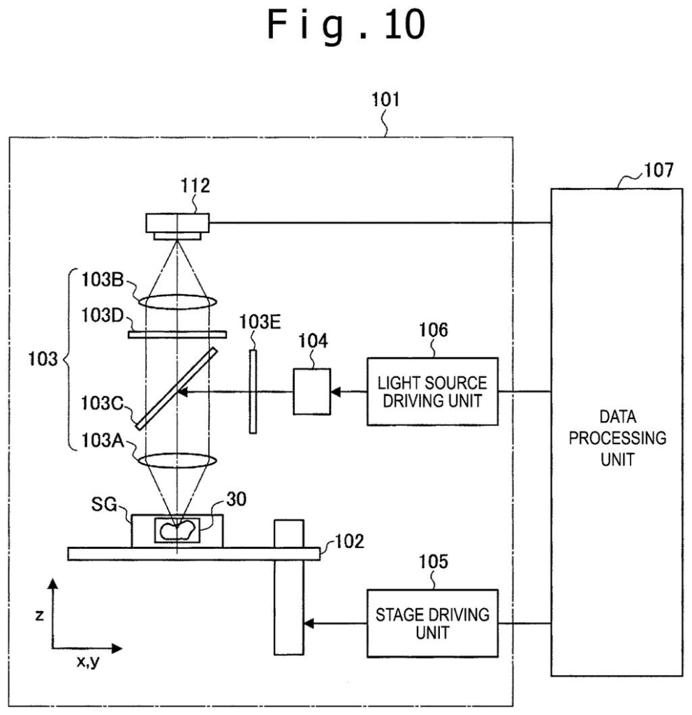
What Sony's stained-vs-unstained cell test actually does
You're a lab technician looking at a slide of cells under a microscope, trying to figure out whether a suspicious cell is actually dangerous or just looks that way because of the chemical dye you used to make it visible. That dye can sometimes create false signals, and right now there's no easy automated way to subtract that noise.
Sony's patent describes a system that runs the same computer analysis twice: once on a dyed sample and once on an identical undyed sample. If the dyed scan flags a cell as abnormal but the undyed scan doesn't, the system knows the dye itself might be responsible. By comparing the two results, it produces a score indicating whether the cell is a genuine positive (truly abnormal) or a false alarm.
The goal is to make automated cell screening more trustworthy, reducing the chance that a staining artifact, rather than a real biological signal, drives a diagnosis. That matters most in medical contexts where a missed or wrong result carries serious consequences.
… a stained specimen prepared by staining a specimen containing a target cell with a fluorescent reagent …
Translation: A tissue sample is treated with a glowing dye to highlight specific cells.
How the processor compares two scans to flag positive cells
The patent describes a two-step image analysis pipeline designed to improve the accuracy of positive cell determination, the process of deciding whether a cell in a biological sample carries a meaningful biological marker.
First, a processor runs first analysis processing on a stained image: a photograph of tissue that has been treated with a fluorescent reagent (a dye that makes certain molecules glow under specific lighting). That analysis includes automated cell detection and any further classification steps.
Second, the processor runs the exact same analysis on an unstained image of a specimen that is the same as, or closely similar to, the stained one. Running identical processing on both versions is the core design choice: if the algorithm finds something in the stained image that it doesn't find in the unstained image, that difference can be attributed to the dye rather than to a real biological feature.
Finally, the system outputs an index (a numerical or categorical score) based on the gap between the two results. A large difference suggests the stain is genuinely lighting up a real target cell; a small difference suggests the stain may be creating a spurious signal. The claim covers the method broadly, without specifying a particular cell type, staining reagent, or downstream AI architecture.
… outputting an index indicating whether or not the target cell is a positive cell, based on a difference between an analysis result of the first analysis processing and an analysis result of the second analysis processing …
Translation: The system calculates a final score by comparing the computer scans of the dyed and undyed tissue.
What this means for lab testing and cell analysis tools
For anyone whose medical care depends on cell-level lab tests, accuracy gaps in automated screening are a real concern. False positives from staining artifacts can trigger unnecessary follow-up procedures, while false negatives can delay treatment. A system that actively cross-checks a stained result against an unstained baseline adds a layer of verification that purely single-image analysis lacks.
Sony's interest in medical imaging and analysis puts this filing in a broader context: the company has hardware and sensor expertise that could make it a supplier of both the imaging equipment and the analysis software for clinical labs. Whether this method reaches a product or gets licensed to diagnostics companies, the underlying claim is broad enough to cover a wide range of cell types and staining protocols.
Sony's 540th filing in our Sony coverage since May adds to a run that includes a GPS accuracy fix and smoother AR lens edges.
Claim 1 as written is broad. It covers any method that runs the same analysis on a stained image and an unstained image of a matching specimen, then outputs any index based on the difference. There is no limitation on the type of cell, the type of fluorescent dye, the specific detection algorithm, or the form the index takes. That breadth is a double-edged situation for Sony.
On one hand, a broad claim, if granted, could give Sony a wide enforcement perimeter over any automated lab system that uses this compare-stained-to-unstained design, even if competitors implement the underlying math very differently. On the other hand, broad claims are also the ones most likely to attract prior-art challenges during examination, because the idea of using a control image (an unstained reference) to correct for staining artifacts is not conceptually new in biology.
The interesting question the USPTO examiner will face is whether the specific framing here, running identical algorithmic processing on both images and outputting a difference-based index, is distinct enough from existing image subtraction and background-correction techniques in digital pathology. If Sony can hold the claim as written, it is a meaningful position in a growing automated diagnostics market. If the claim gets narrowed, it may end up protecting only a specific implementation.
There are more where this came from
We read every patent application Big Tech publishes and send you the ones worth knowing. Plain English, free, every week.
The drawings
26 drawing sheets from US 2026/0287508 A1 · click any drawing to enlarge


Want this weekly breakdown for a company we don't cover? Patentlyze Pro →
Be the first to weigh in